

Ventral Bulla Osteotomy
What is a ventral bulla osteotomy?
A ventral bulla osteotomy or VBO for short is a surgical procedure to access the middle ear and is most commonly performed in cats to remove nasopharyngeal polyps. The bulla is a boney structure at the base of the skull that houses the structures of the middle ear.

Nasopharyngeal polyps are growths that occur in the middle ear and can extend out the ear canal or into the nasopharyngeal region through the eustachian tube.
What are signs of a nasopharyngeal polyp?
Nasopharyngeal polyps occur most frequently in young cats and signs include:
Discharge from the ear canal
A pink fleshy growth in the ear canal
Nasal discharge
Stuffy nose or difficulty breathing
Nasopharyngeal polyps occur secondary to chronic inflammation, likely related to upper respiratory tract viruses in kittens.
Nasopharyngeal polyps can sometimes be diagnosed on physical exam, but often your vet will recommend a CT scan of the head to more fully evaluate the middle ear and nasopharynx region.
Figure 1: View of the skull of a cat from below.

Figure 2: CT scan image of a cat skull showing the tympanic bulla filled with a polyp.
What to expect during the procedure:
If a large mass is found exiting into the ear canal, the mass may need to be pulled from the ear while your cat is under general anesthesia. If the mass is protruding into the nasopharynx, the mass may be pulled out through the mouth. After removal of the mass, any remaining tissue will be removed from middle ear by a ventral bulla osteotomy (VBO) procedure. During the VBO, an incision is made under the jaw to access the bulla. An opening is made into the bulla to allow removal of the diseased tissue and debris. The bulla is flushed and any remaining tissue removed. The tissue removed will be sent for biopsy and culture.
After surgery, your pet will be sent home with antibiotics and pain medications. It is important to restrict activity for 10-14 days after surgery while the incision heals. Monitor the incision for signs of redness, swelling or discharge and do not allow your pet to rub or scratch at the incision. An Elizabethan collar may be used to prevent self-trauma. It may be hard for your cat to eat hard food after surgery, so canned food is recommended for the first 3-5 days.
What to expect after surgery:
Most cats do very well after surgery. In cases where the polyp was occluding the nasopharynx, immediate improvement in breathing is to be expected.
Possible complications associated with a ventral bulla osteomy include:
Bleeding
Swelling of the neck region
Vestibular signs (vertigo, head tilt, dizziness)
If these signs occur, they are typically temporary.
Recurrence of the polyp
Horner’s Syndrome.
Horner’s Syndrome is due to damage to the sympathetic nerve fibers that are present in the middle ear. It is expected to see Horner’s Syndrome after a VBO and in most cases this is temporary, resolving during the first few weeks after surgery. Dogs and cats with Horner’s Syndrome will have elevation of the third eyelid in the affected eye as well as drooping of the eyelid and constriction of the pupil. These changes do not cause pain or distress and do not require any treatment.
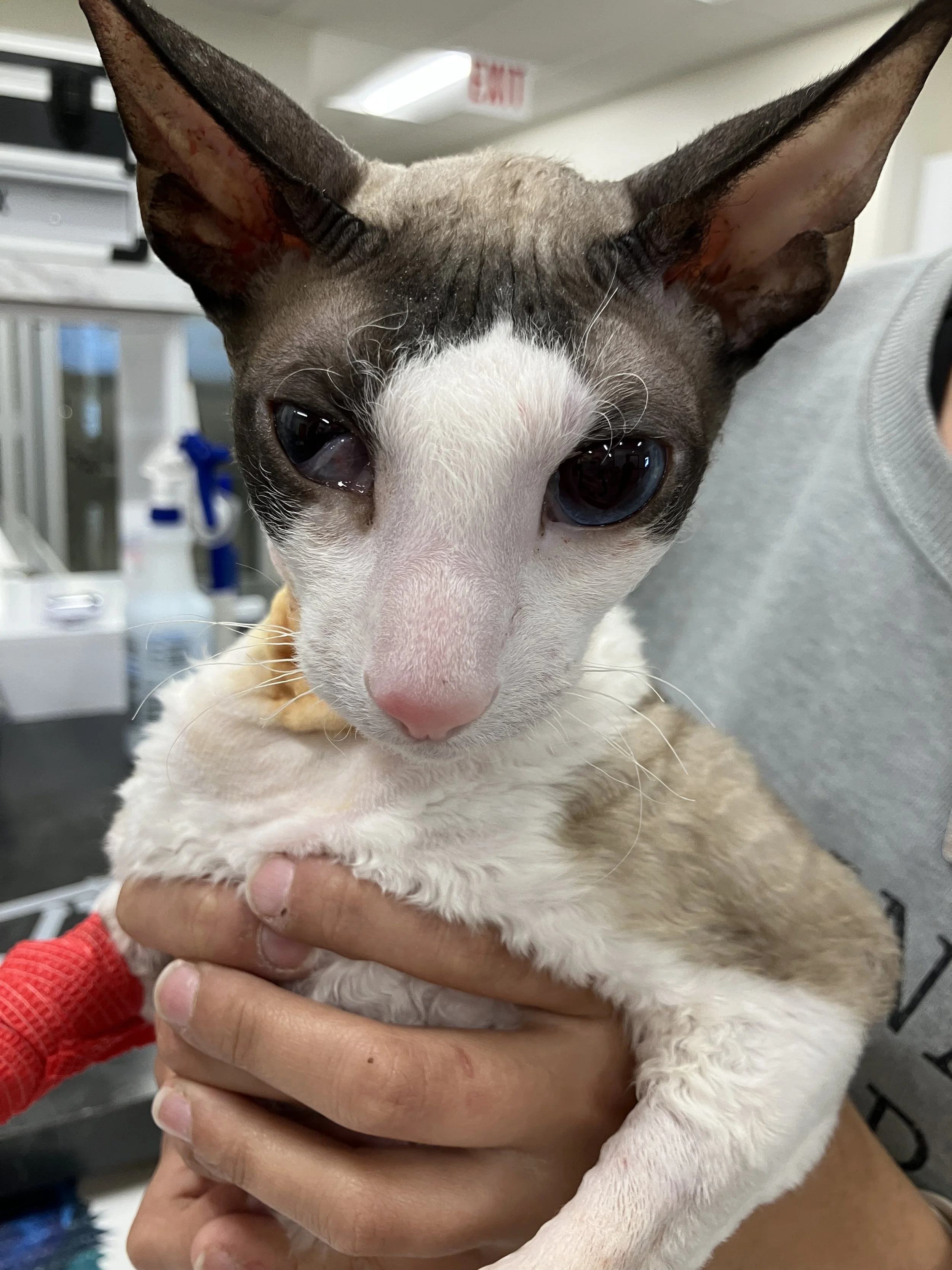
Figure 3: Horner’s Syndrome in the right eye of a cat after a VBO.
What is recovery like?
After surgery your pet will need to have activity limited for about 10-14 days and the incision site kept dry. No running, jumping or playing should be allowed. The incision should be monitored for any redness, swelling or discharge and an E-collar should be used to prevent your pet from scratching or rubbing the incision. Pain medications will be prescribed to be given for several days after surgery and antibiotics may be used if appropriate.
Immediately post-surgery, your pet may be drowsy, uncoordinated or nauseous. Unless otherwise instructed, we normally recommend the following for food and water:
Water Reintroduction: Offer a small amount of water 30 minutes to 1 hour after arriving. If there are no signs of nausea and water is kept down, more can be offered in small amounts. You may resume normal water access the following day,
Food Reintroduction: Offer 1/2 their normal feed 2 hours after arriving home. If there are no signs of nausea and food is kept down, you may resume normal feedings the following day.
Detailed postoperative instructions will be provided to you after the surgery that outline medications, and incision care. Bandages on the IV catheter site can be removed once you get home. It is normal for some bruising and swelling to occur. If there is no pain or discharge associated with the swelling, continue to monitor at home. Please notify us or your veterinarian if you observe:
Increased redness/bruising over time
Odorous or pus-like discharge
Opening of the incision site
Increased swelling
Vomiting
Signs of acute pain.