
Total Ear Canal Ablation and Bulla Osteotomy
An incision is made around the opening of the ear, and the entire ear canal is removed. The tympanic bulla is opened, and the inside lining is removed. A culture of the bulla is taken at surgery to determine what antibiotics will be needed. The pinna or ear flap is left in place.
The affected ear becomes nonfunctional after surgery; however, most pets already have significant hearing loss prior to the procedure due to ear disease.
Potential Complications
Like all surgeries, TECA+BO carries some risks including:
Facial nerve paralysis: The facial nerve travels behind the ear canal and can be damaged during dissection of the ear canal or may become traumatized due to swelling and inflammation resulting in dysfunction in up to 50% of cases. Facial nerve paralysis results in the inability to blink the eyelid on the affected side. Facial nerve paralysis may be partial or complete and may be permanent, or more commonly, temporary. Temporary dysfunction will correct itself over a 2–4-week period after surgery.
Dogs and cats with facial nerve dysfunction quickly learn to use the third eyelid to “blink” their eye and keep it lubricated. In the interim, eye lubricant medications should be used to prevent damage to the cornea.
Horner’s syndrome: Drooping of the eyelid, a small pupil and elevation of the third eyelid. This occurs due to irritation of the sympathetic nerves within the middle ear and is more common in cats. Horner’s syndrome does not typically result in functional issues.
Infection: Occasionally, an abscess will develop in the region of the ear. This can occur even years after surgery and signs include pain on opening of the jaw or the development of an open, draining tract. An infection develops when tissue is retained within the tympanic bulla and surgery is often necessary to remove the tissue.
Bleeding: Due to the presence of multiple veins and arteries in the vicinity of the ear canal and bulla, serious bleeding can occur during the surgical procedure. Bleeding after surgery is rare.
Because of the risks associated with performing bilateral (both right and left side) TECA+BOs at the same time, it is recommended to perform surgery on one ear, then allow several weeks of healing to occur before performing a TECA+BO on the other ear for pets that need both removed.
What is recovery like?
After surgery your pet will need to have activity limited for about 10-14 days and the incision site kept dry. No running, jumping or playing should be allowed. The incision should be monitored for any redness, swelling or discharge and an E-collar should be used to prevent your pet from scratching or rubbing the incision. Pain medications will be prescribed to be given for several days after surgery and antibiotics are used to treat any residual infection from deep within the ear canal. The antibiotic may need to be changed based on the culture results, which should be complete within about a week after surgery. Antibiotics are continued for several weeks after surgery.
Immediately post-surgery, your pet may be drowsy, uncoordinated or nauseous. Unless otherwise instructed, we normally recommend the following for food and water:
Water Reintroduction: Offer a small amount of water 30 minutes to 1 hour after arriving. If there are no signs of nausea and water is kept down, more can be offered in small amounts. You may resume normal water access the following day,
Food Reintroduction: Offer 1/2 their normal feed 2 hours after arriving home. If there are no signs of nausea and food is kept down, you may resume normal feedings the following day.
Detailed postoperative instructions will be provided to you after the surgery that outline medications, and incision care. Bandages on the IV catheter site can be removed once you get home. It is normal for some bruising and swelling to occur. If there is no pain or discharge associated with the swelling, continue to monitor at home. Please notify us or your veterinarian if you observe:
Increased redness/bruising over time
Odorous or pus-like discharge
Opening of the incision site
Increased swelling
Vomiting
Signs of acute pain.
The cosmetic results after TECA+BO are quite good and often the only way to tell that surgery has been performed is to lift the ear flap and note that there is no longer an opening into the ear canal. In dogs that have erect ears, the ear pinna may slightly droop.
Why is a TECA+BO performed?
A total ear canal ablation or TECA is a surgical procedure performed to treat severe, end stage ear disease.
This procedure is indicated for dogs and cats with:
Chronic ear infections that have failed to respond to medical treatment. Infections are typically related to underlying allergies in dogs and cats.
Tumors or growths in the ear canal
Failure of the ear canal to properly develop
Trauma to the ear canal
A TECA+BO is a salvage procedure, performed when medical therapy has failed or is not indicated. This procedure can result in significant pain relief and improved quality of life.
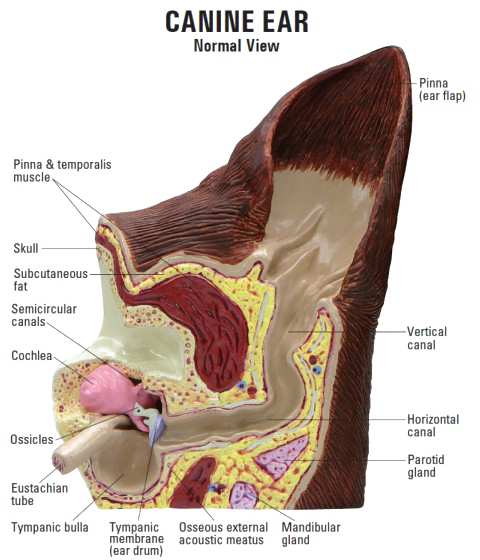
During this procedure, the entire ear canal is removed (total ear canal ablation) and the middle ear cavity is opened (bulla osteotomy) to remove all diseased tissues. The flap of the ear is called the pinna. The ear canal of the dog and cat is a cartilaginous tube, shaped like an “L”. The tympanic bulla is the boney cavity of the skull where the ear canal attaches.
Please note, for surgery, the fur around the ear will be shaved down and your pet’s face will look different without it. The fur will grow back!
Prognosis
Dogs and cats are expected to experience significant improvements in their quality of life after healing from a TECA+BO. Pets who under the procedures for both sides will completely lose their hearing but they will adapt and can be taught to respond to visual cues.
Because the ear canal has been removed, there is no need for continued ear cleaning and medicating in pets affected by allergies.