Mast Cell Tumor Removal
What is a Mast Cell Tumor?
Mast cell tumors (MCTs) are the most common type of skin tumor found in dogs and represent about 20% of all skin tumors. These tumors arise from the mast cell, which is a type of cell involved in the allergic response. MCTs can be limited to the skin or they can spread to other parts of the body including the lymph nodes, the spleen, liver or bone marrow. These tumors are often solitary, but about 10% of pets will develop MCTs in multiple locations.
What do they look like?
Mast cell tumors can look like anything and are often referred to as the “great imposter”. Some are small, raised lumps, while others may be red, ulcerated or itchy. MCTs can also vary in size on a daily basis due inflammation caused by substances (histamine granules) within the mast cells themselves.
Common locations for MCTs:
Limbs 24-40%
Groin or genital area
Face 10-14%
Body (trunk) 50-60%
Because of their ability to look like anything, it is recommended that any lump or bump be evaluated by your veterinarian.
Most mast cell tumors do not make your dog or cat sick; however, some pets will develop lack of appetite, vomiting or melena (bloody stools) due to gastrointestinal bleeding secondary to the release of factors from the mast cells.
How are MCTs diagnosed?
Most MCTs can be diagnosed by performing a fine needle aspirate (FNA). To perform this test, your veterinarian will insert a small needle into the tumor to remove some of the cells. They will then look at these cells under a microscope. Mast cells appear as round cells that contain dark purple dots, or granules.
Staging and Grading
Staging refers to tests that are performed to try to determine the extent of mast cell disease in your pet’s body.
Limited staging includes the following:
Aspiration of the local lymph node(s)
Complete staging includes the following:
Aspiration of the local lymph node(s)
Thoracic radiographs
Abdominal ultrasound
Aspirates of the spleen and liver
+/- removal of the local lymph node
The decision to do limited or complete staging is influenced by multiple factors including the location or suspected aggressiveness of the tumor, resources and personal preferences. These decisions can be made in conjunction with your surgeon.
The grade of the tumor refers to how aggressive the tumor is expected to behave. The grade is based on microscopic features of the tumor such as invasiveness into the surrounding tissues and the number of actively dividing cells. In general, mast cell tumors can be divided into 3 grades (I, II, III) or more recently into low- and high-grade tumors. High grade tumors tend to be more aggressive and more likely to spread while low grade tumors are less likely to spread.
What is the Treatment for a MCT?
Treatment options depend on several factors including the location of the tumor and if there is any evidence of spread of the disease. Surgical removal is the preferred treatment when possible. Before surgery, an antihistamine such as Benadryl to lessen the chances of the mast cells releasing histamine from their granules and causing inflammation is recommended as well as an antacid such as PepcidAC or Prilosec as pets with MCTs often have high levels of acid in their stomach. Please always confirm with your veterinarian before administering any medication.
Mast cell tumors invade into the surrounding tissues so wide surgical excision (removal of not only the tumor but a wide are of healthy tissue surrounding the tumor) is necessary to completely remove the disease. This often results in an incision that is significantly larger than the original tumor.
After surgery, the mass will be sent to the laboratory for a veterinary pathologist to evaluate. They will give us information such as whether the entire tumor was able to be removed as well as the grade of the tumor. This information if used to help determine a prognosis and if additional therapy is needed.
What is recovery like?
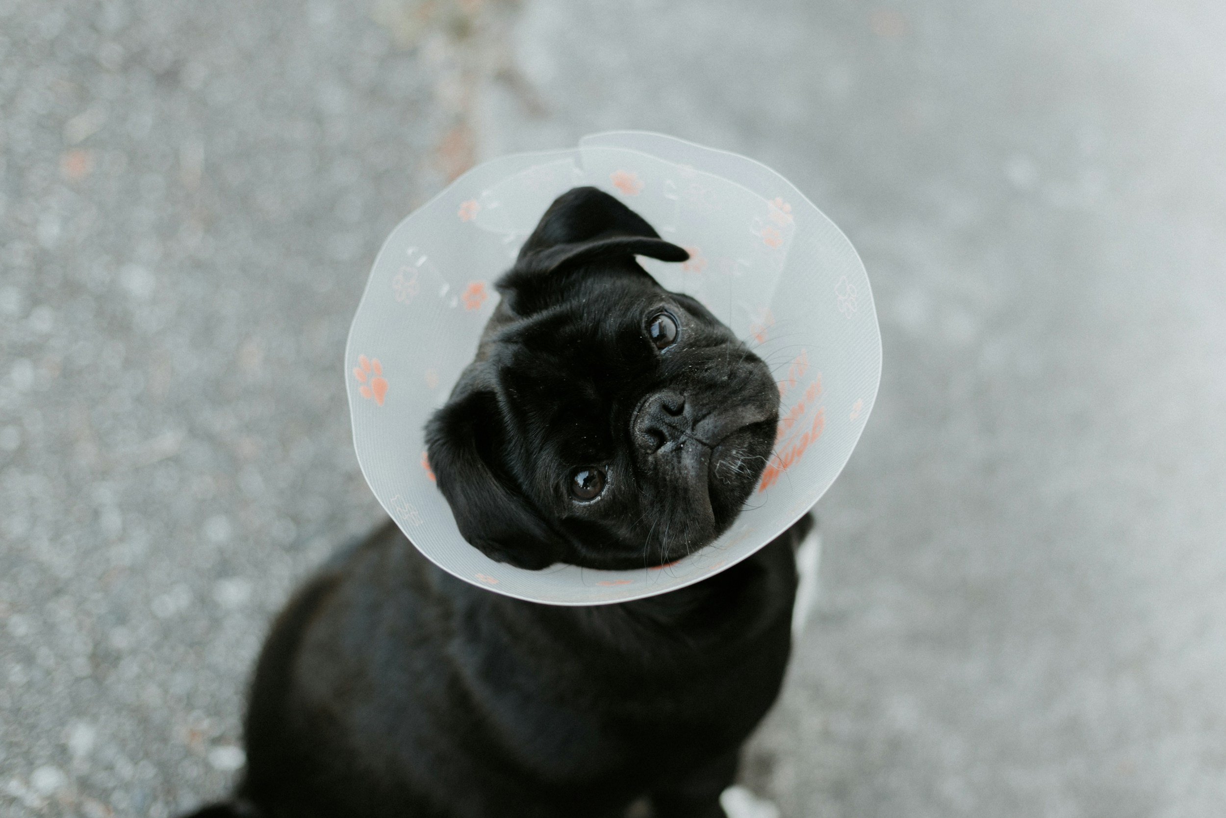
After surgery your pet will need to have restricted activity for 10-14 days while the incision heals. No running, jumping or playing should be allowed and they should stay on a leash when taken outside to use the bathroom. Your pet will also be sent home with pain medications to help keep them comfortable. An E-collar (a protective device) is often necessary to prevent any licking or chewing of the incision.
The incision should be monitored for any redness, swelling or discharge.
We recommend continuing the antihistamine and antacid for about a week after surgery, but please confirm with your veterinarian. Promptly report any symptoms like vomiting, diarrhea, appetite loss or behavior changes to us or your veterinarian.
Immediately post-surgery, your pet may be drowsy, uncoordinated or nauseous. Unless otherwise instructed, we normally recommend the following for food and water:
Water Reintroduction: Offer a small amount of water 30 minutes to 1 hour after arriving. If there are no signs of nausea and water is kept down, more can be offered in small amounts. You may resume normal water access the following day,
Food Reintroduction: Offer 1/2 their normal feed 2 hours after arriving home. If there are no signs of nausea and food is kept down, you may resume normal feedings the following day.
Detailed postoperative instructions will be provided to you after the surgery that outline medications, and incision care. Bandages on the IV catheter site can be removed once you get home. Local bandages applied to the surgical site can be removed in 72 hours unless otherwise instructed. It is normal for some bruising and swelling to occur. If there is no pain or discharge associated with the swelling, continue to monitor at home. Please notify us or your veterinarian if you observe:
Increased redness/bruising over time
Odorous or pus-like discharge
Opening of the incision site
Increased swelling
Prognosis
Complete surgical removal of low-grade tumors is generally curative, and no additional treatment is necessary. Your vet may recommend a consultation with a veterinary oncologist to discuss chemotherapy or radiation therapy for higher grade tumors, tumors that are incompletely removed or for tumors in certain parts of the body (face and groin) that may behave more aggressively.
Some dogs that have a low grade MCT removed may develop a new "“de novo” MCT months to years later, so it is always important to keep monitoring your pet for any new lumps and bumps.
Many dogs live happy, full lives after treatment for MCTs. Early diagnosis and appropriate management are key. If you have any questions, we are here to guide you every step of the way.
